Swept Confocally-Aligned Planar Excitation (SCAPE 2.0)
Swept, Confocally-Aligned Planar Excitation (SCAPE) microscopy is a technique for high-speed 3D microscopy in living organisms. SCAPE can image intact living samples including the intact mouse brain and freely moving organisms such as Drosophila melanogaster larvae and the zebrafish heart at up to 300 volumes per second. We originally introduced SCAPE in 2015, published in Nature Photonics. Our new paper in Nature Methods describes and demonstates SCAPE 2.0, our new and improved, even faster, even higher resolution version of the system. SCAPE is licensed to Leica Microsystems for commercial development.
Our lab is working on many aspects of SCAPE, including developing more systems that can reach wider applications, including higher resolution, larger fields of view, and deeper imaging into scattering tissues using two-photon excitation. We are also working with a wide range of collaborators to apply SCAPE to specific research applications. For details on applications and collaborations see HERE. We are also sharing SCAPE - helping labs around build their own versions of the system to accelerate their biomedical research. For more information on 'Open-SCAPE' see HERE.
SCAPE is a hybrid between light-sheet microscopy and confocal scanning microscopy that overcomes many of the limitations of these existing technologies:
1) SCAPE uses a single objective lens for both illumination and detection, making sample positioning and alignment much simpler than conventional light-sheet imaging.
2) SCAPE uses an oblique light sheet which is swept through the sample using a scanning mirror, capturing images of the optically sectioned illuminated plane as it sweeps. This means that SCAPE acquires 3D volumetric images without needing to physically translate the objective lens or the sample. This feature significantly increases achievable imaging speeds, while avoiding complex sample preparation and enabling greater sample diversity.
3) SCAPE's unique scanning and de-scanning optics mean that the illuminated plane always stays aligned with a stationary camera providing optical sectioning with no other moving parts. When bidirectional scanning, there is no duty cycle or overhead resulting in a simple, inexpensive system with very high volumetric imaging speeds.
4) SCAPE resolution rivals light-sheet, confocal and two-photon microscopy in living tissues. Our current prototype uses an inexpensive 488 nm CW for illumination and in transparent samples and phantoms can image fields of view 1 mm wide by 600 microns deep. Penetration is more limited in scattering tissues such as brain (as is also the case in conventional light-sheet microscopy). However, we have already demonstrated imaging of spontaneous GCaMP activity apical dendrites of layer 5 neurons within layers 1 and 2 of the rodent cortex. We expect future implementations of SCAPE to improve penetration depth significantly.
SCAPE System Design
The imaging geometry of SCAPE is shown below. The sequence of images depicts how a 3D volumetric image is formed as the oblique light sheet sweeps through the sample.
Additional optical components in our system take the stationary oblique image plane and map it onto the surface of a high-speed camera. We use and Andor Zyla 5.5 camera which can read out 80 rows (and 2,560 columns) of its camera chip at 2,400 frames per second. In this configuration, the result would be image volumes with 80 depths (z'), 2,560 voxels in y' and 200 voxels in x' at 12 volumes per second. For 100 voxels in x' the volume rate would be 24 VPS. Faster cameras could provide rates exceeding 100 VPS. All data shown to date has been acquired using a 30 mW or 50 mW 488 nm DPSS CW laser.
Imaging the Living Brain
The unique, single, stationary objective configuration of SCAPE mean that it can be used just like any other upright or inverted microscope. Where the conventional two-objective geometry of light-sheet microscopy has prevented its use for imaging the intact brain, SCAPE can achieve high-speed, optically sectioned imaging of the superficial layers of the cortex. Two-photon microscopy of the brain is limited in 3D imaging speed owing to the need to visit every point in the sample with a single scanning spot of light. SCAPE can image equivalent volumes at around 100 times faster than standard two-photon microscopy. Unlike random-access two-photon scanning, SCAPE requires no a-priori information or selection of regions of interest, making it possible to capture spontaneous dynamics of GCaMP in dendritic branches in the superficial layers of the cortex, as shown below. Temporal resolution and signal to noise are sufficient to examine the onset and decay dynamics of GCaMP transients for a single spontaneous event, spatially resolved within a single dendritic tree. See Bouchard et al for full details.
The movie below shows a dual-color SCAPE volume showing both blood flow and neural activity acquired in the awake, behaving brain at 10 volumes per second. Data was acquired in collaboration with Randy Bruno and Clay Lacefield.
Imaging Freely Moving Organisms
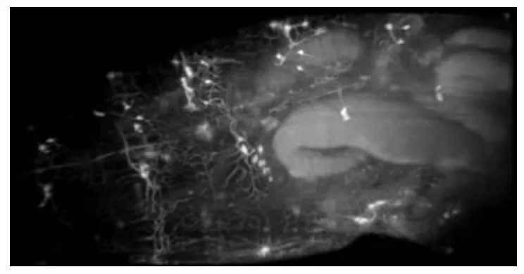
Understanding the nervous system is to understand the activity of neurons during behaviour. A major goal of the BRAIN Initiative is to capture functional actvity within the whole brain, or even whole body of complete organisms. Drosophila melanogaster , fruit flies, are a commonly used model organism in neuroscience. SCAPE has the ideal field of view and volumetric imaging speed to capture the whole body of freely moving fruit fly larvae, permitting us to asses not just 3D motion and structure, but the function of cells during this behavior. The video embedded above / below shows a sequence of different SCAPE data sets acquired in a range of transgenic Drosophila models expressing GFP and GCaMP. These data were acquired in collaboration with Richard Mann, Cesar Mendes and Wes Grueber.